Fig. 5.
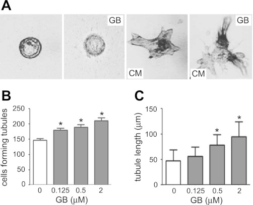
GB promotes the tubulogenesis in MDCK cells and MDCK cysts. A: representative light micrographs of tubule-like structures induced from the MDCK cysts in collagen gels. Light micrographs were taken on day 12 after MDCK cysts (established on day 4 with FSK stimulation) cultured without 3T3 conditioned media (CM; left) or with GB and without 3T3 CM (second from left) or with 3T3 CM (third from left), or with 3T3 CM and GB (right). Scale bar = 100 μm. B: numbers of MDCK cells forming tubule-like structures without or with GB treatment (means ± SD; n = 3). *P < 0.05 vs. control. C: average values of the longest length of tubule-like structures on each 3T3 CM-treated MDCK cyst without or with GB treatment (means ± SD; n = >30). *P < 0.05 vs. control.

