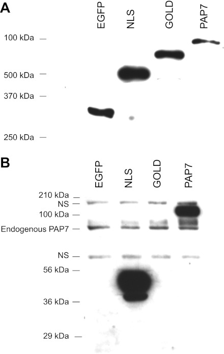
Fig. 3.
Confirmation of the specificity of the anti-PAP7 antiserum. K562 cells were transfected with the various enhanced green fluorescent protein (EGFP)-PAP7 constructs EGFP vector alone, EGFP-nuclear localization signal (NLS), EGFP-Golgi dynamics domain (GOLD), and EGFP-PAP7 as described in the materials and methods. At day 1 after transfection cell lysates were prepared, proteins separated by SDS-PAGE and Western blots performed with anti-GFP antibody (A) or anti-PAP7 antiserum (B). NS refers to nonspecific bands that were always present with the anti-PAP7 antiserum. Molecular weight (MW) markers are also shown for A and B as is the endogenous PAP7 present in the K562 cells.

