Fig. 4.
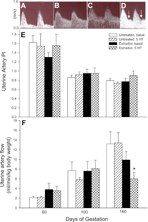
A–D: representative uterine artery flow waveforms as assessed by Doppler ultrasonography on day 60 (A) and day 160 (B) during saline infusion (i.e., basal) in untreated baboons and on day 160 in animals treated with estradiol on days 25–59 and during infusion of saline (C) or 5-HT (8 μg·min−1·kg body wt−1; D). E and F: mean ± SE values of uterine artery pulsatility index (PI; E) and uterine artery volume flow (in ml·min−1·kg maternal body wt−1; F) as assessed on days 60, 100, and 160 of gestation in untreated (n = 5) and estradiol-treated (n = 6) baboons during saline (basal) or 5 HT (8 μg·min−1·kg body wt−1) infusion. Uterine artery PI values for untreated and estradiol-treated animals during the infusion of saline or 5-HT on days 100 and 160 were lower (P < 0.01) than the respective values on day 60. A progressive increase (P < 0.01) in volume flow occurred in untreated animals in the basal state between days 60, 100, and 160. An increase (P < 0.05) in volume flow in estradiol-treated animals in the basal state occurred between days 60 and 100 only. Arrows in D designate notching. *P < 0.01 vs. untreated 5-HT-infused animals and P < 0.05 vs. estradiol-treated animals in the basal state.

