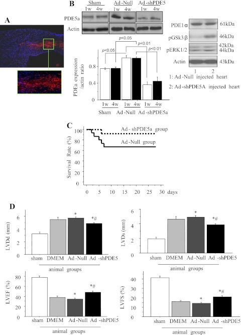
Fig. 1.
Expression of phosphodiesterase-5a (PDE5a) in vivo, survival curves, and left ventricular (LV) heart function in various treatment groups of animals. A: a typical injection site in adenoviral vector encoding for short hairpin RNA sequence targeting PDE5a (Ad-shPDE5a)-injected animal heart in the peri-infarct region identified by DS Red (red fluorescence) on day 7 after treatment. 4′,6-Diamidino-2-phenylindole (DAPI) was used to visualize the nuclei (original magnification ×10). Area in the green box has been magnified for clarity. B: Western blot showing PDE5a expression in the LV of infarcted hearts at 1 and 4 wk after treatment with adenoviral vector without therapeutic shRNA (Ad-Null) or Ad-shPDE5a using sham-operated animals as a baseline control. Graph shows PDE5a expression increased significantly in Ad-Null-treated infarcted hearts compared with sham-operated heart. Ad-shPDE5a abrogated PDE5a expression both at 1 wk and 4 wk after treatment (P < 0.01 vs. both Ad-Null and sham-operated animals). No change in PDE1a was observed in the animal hearts with their respective treatment. C: survival curves from Ad-Null and Ad-shPDE5a treatment groups of animals. Compared with 67% in the Ad-Null-treated group, the survival rate was 92% in the Ad-shPDE5a-treated group (P = 0.11) D: echocardiographic data for LV geometry and function 4 wk posttreatment with Ad-Null and Ad-shPDE5a. LV diastolic diameter (LVDd), LV end-systolic diameter (LVDs), LV ejection fraction (LVEF), and LV fractional shortening (LVFS) were significantly preserved in Ad-shPDE5a-treated animal hearts. Values are means ± SE. *P < 0.05 vs. DMEM-treated mice; #P < 0.05 vs. sham-operated mice.

