Fig. 3.
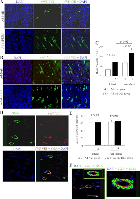
Blood vessel density and maturation index in infarct and peri-infarct regions of heart from different treatment groups. Photomicrographs showing von Willebrand factor (vWF)-positive vessels (green) in infarct (A) and peri-infarct areas (B) at 4 wk after treatment. DAPI, 4′,6-diamidino-2-phenylindole. C: graph comparing the number of vWF-positive blood vessels between groups. *P < 0.05 vs. Ad-Null-treated mice in infarct area; #P < 0.05 vs. Ad-Null-treated mice in peri-infarct area. HPF, high-power field (×400). D, E: representative photomicrographs of tissue sections from Ad-shPDE5a-treated heart immunolabeled for vWF-VIII (red) and α-smooth muscle actin (SMA, green). Graph for blood vessel maturation index showing insignificant change in maturation index between Ad-shPDE5a- and Ad-Null-treated animal hearts in both infarct and peri-infarct regions. Values in the graph are means ± SE. F: double fluorescence immunostaining of the LV in the normal myocardium for vWF-VIII (red) and α-SMA (green) expression showing a typical blood vessel depicting endothelial (red) and SMA (green) staining (original magnification ×20).

