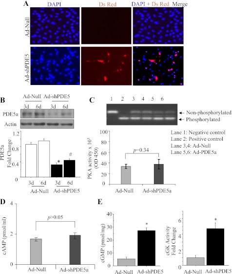
Fig. 5.
Genetic manipulation of cardiomyocytes with Ad-shPDE5a. A: the cultured cardiomyocytes transduced with Ad-shPDE5a (DsRed positive, red fluorescence) at 72 h after transduction. B: Western blot showing successful abrogation of PDE5a expression in cultured cardiomyocytes observed on day 3 and day 6 after transduction with Ad-ShPDE5a. Values plotted in the graph are means ± SE. *P < 0.05 vs. Ad-Null-treated group on day 3; #P < 0.05 vs. Ad-Null-treated group on day 6. C, D: activity assays showing insignificantly increased PKA and cAMP activity in the cardiomyocytes transduced with Ad-shPDE5a compared with Ad-Null treated cardiomyocytes. E: cGMP and PKG activity assays in cultured cardiomyocytes on day 3 after transduction with Ad-shPDE5a. Values are means ± SE. *P < 0.05 vs. Ad-Null-treated group.

