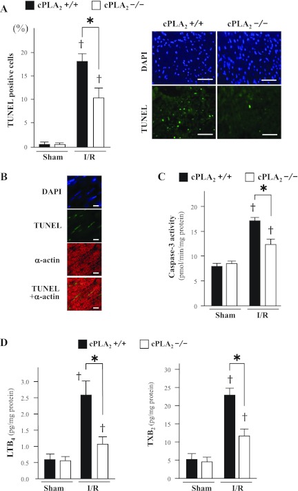
Fig. 4.
dUTP nick end-labeling (TUNEL) assay, caspase activity, and leukotriene B4 (LTB4) and thromboxane B2 (TXB2) content in myocardium. A: left, percentage of TUNEL-positive nuclei in sham-operated myocardium (Sham) and in noninfarcted myocardium in the ischemic area after 24 h reperfusion following 1 h ischemia (I/R). The TUNEL-positive nuclei were counted in 6 fields of 5 separate sections at ×400 magnification and are presented as a percentage of the total number of cardiomyocytes (nuclei). Right, representative TUNEL staining in noninfarcted myocardium in the ischemic area after I/R. TUNEL-positive nuclei are stained green [fluorescein isothiocyanate (FITC)]. The entire cell population was visualized under a fluorescence microscope with a DAPI filter. Scale bars = 50 μm. B: representative immunofluorescence microscopic images of ischemic myocardium of cPLA2α+/+ mice stained for TUNEL (green) and α-actin (red). Scale bars = 10 μm. C: caspase-3 activity in sham-operated myocardium and in noninfarcted myocardium in the ischemic area after I/R. D: content of LTB4 and TXB2 in homogenates of noninfarcted myocardium in the ischemic area after I/R. *P < 0.05 and †P < 0.01 compared with the respective sham-operated mice (n = 6∼12 mice in each experiment).

