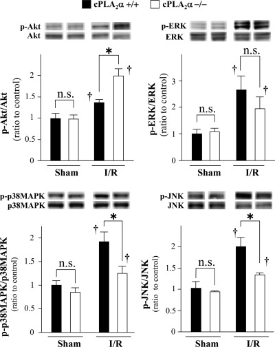
Fig. 6.
Comparison of phosphorylation status of protein kinase B (AKT), ERK1/2, p38 MAPK, and c-Jun NH2-terminal kinase (JNK) in the ischemic myocardium between cPLA2α+/+ and cPLA2α−/− mice. Hearts from cPLA2α+/+ mice (filled bars) and cPLA2α−/− mice (open bars) were harvested at 6 h reperfusion following 1 h ischemia. The expression of total and phosphorylated (p) kinases was assessed by immunoblotting. Insets on top show representative immunoblots. Levels of phosphorylated kinases were expressed as a ratio of the intensity of the respective total protein band and normalized to the sham-operated cPLA2α+/+ myocardium (=1); n = 6 in each experiment. *P < 0.05 and †P < 0.05 compared with the respective sham-operated mice. ns, Not statistically significant.

