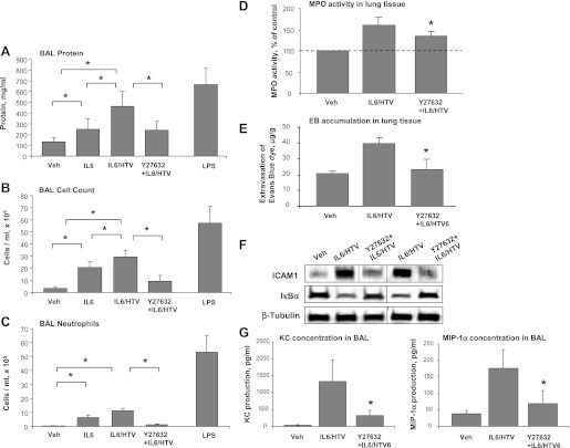
Fig. 7.
Role of Rho in the development of lung injury in IL-6/HTV two-hit in vivo model. Mice were subjected to intravenous injection with vehicle or IL-6 (5 mg/kg it) with or without Y-27632 (2 mg/kg iv) instillation followed by mechanical ventilation at high tidal volume (HTV; 30 ml/kg, 4 h). Control animals were allowed to breathe spontaneously. LPS-treated mice (0.63 mg/kg it) served as positive controls. A–C: measurements of protein concentration (A), total cell count (B), and differential PMN count (C) were performed in bronchoalveolar lavage (BAL) fluid taken from control and experimental animals. Data are expressed as means ± SD of 4 independent experiments; n = 6–10 per condition; *P < 0.05. D: myeloperoxidase (MPO) activity was determined in control and treated lung tissue samples. MPO data are expressed as %control ± SD of 4 independent experiments; n = 6–10 per condition; *P < 0.05. E: Evans blue dye (30 ml/kg iv) was injected 2 h before termination of the experiment. Lung vascular permeability was assessed by Evans blue accumulation in the lung tissue. Quantitative analysis of Evans blue labeled albumin extravasation was performed by spectrophotometric analysis of Evans blue extracted from the lung tissue samples; n = 4 per condition; *P < 0.05. F: expression levels of IκBα and ICAM-1 were determined in control and treated lung tissue samples. Equal protein loading in Western blot experiments was confirmed by determination of β-tubulin content in tissue homogenates. Rearranged lanes from the same blot are outlined by vertical dotted line. G: analysis of keratinocyte-derived chemokine (KC) and macrophage inflammatory protein-1α (MIP)-1α levels was performed in BAL samples from control and treated mice by ELISA. Data are expressed as means ± SD of 4 independent experiments; *P < 0.05.

