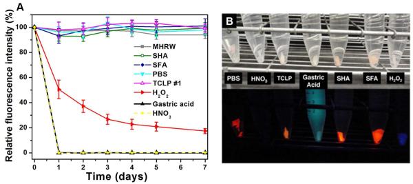
Figure 6.
Fluorescence as a probe for QD damage. (A) Time-resolved relative fluorescence intensity of QD-embedded polymer during fluid exposure. (B) Photographs of QD-embedded polymer after 30-days exposure under visible light (top) and 365 nm UV irradiation (bottom). Note that the light blue color in gastric acid is characteristic of pepsin,36 and the dark blue color of the polymer in H2O2 is possibly the result of light scattering.

