
Figure 6.
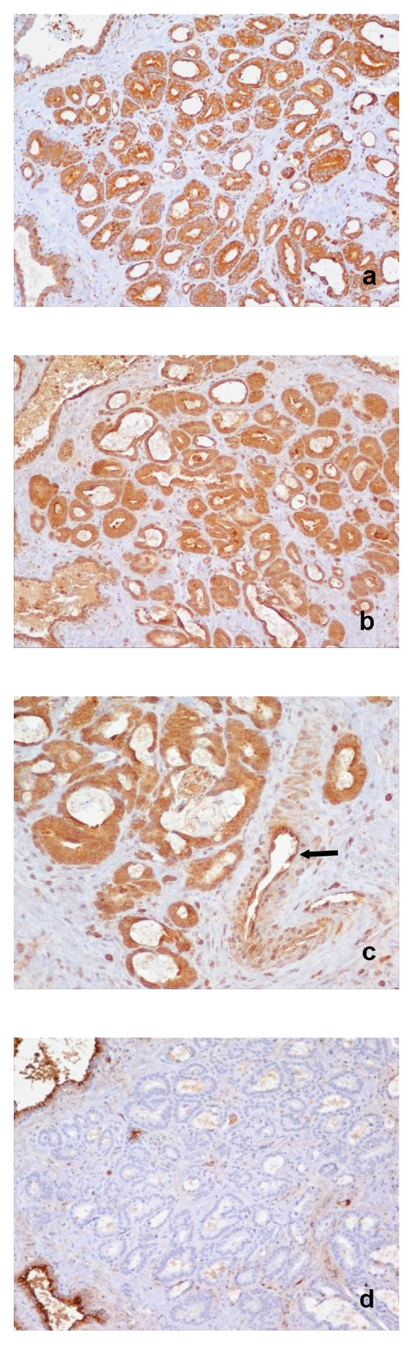
Prostate adenocarcinoma Gleason pattern 3. (a) Strong cytoplasmic IHC staining for NFκB (×200); (b) strong cytoplasmic IHC staining for ET-1 (×200); (c) strong cytoplasmic IHC staining for ET-1. Positive marker the capillary endothelium (arrow) (×400); (d) negative immunoreactivity for NEP. Positive marker the normal prostate glands (×200).