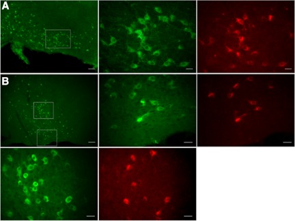
Fig. 1.
Cy3-coupled antibodies raised against mouse p75 neurotrophin receptors (Cy3-p75NTR-IgGs) specifically label cholinergic neurons in the basal forebrain (BF). Photographs show neurons of the horizontal limb of the diagonal band (hDB), magnocellular preoptic nucleus (MCPO), and substantia innominata (SI) nuclei. p75NTR-IgG label is shown in red and choline acetyltransferase (ChAT) immunoreactivity in green. A: ChAT immunostaining in the hDB shown at low magnification (left; scale bar = 100 μm). ChAT-positive neurons and p75NTR-IgG-positive neurons (boxed area) are shown at higher magnification at middle and right, respectively (scale bars = 25 μm). B: ChAT immunostaining in the MCPO and SI at the level of the crossing fibers of the anterior commissure is shown at low magnification (top row, left; scale bar = 200 μm). ChAT-positive neurons and p75NTR-IgG-positive neurons of the MCPO (bottom boxed area) and of the SI (top boxed area) are displayed at higher magnification at top row, middle and right, for MCPO and at bottom row, left and middle, for SI (scale bars = 25 μm).

