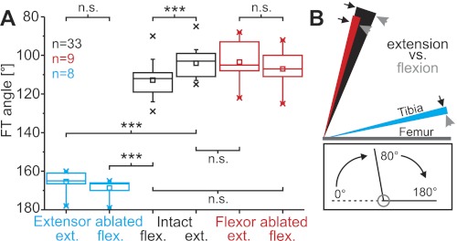
Fig. 1.
A: the hind leg femorotibial (FT) resting angle of intact legs (black) was significantly more flexed after release from full flexion (intact flexed) than after release from full extension (intact extended). Ablation of the extensor tibiae muscle led to a significant increase of the resting angle (tibial flexion) following release from either full extension or full flexion (blue), whereas ablation of the flexor muscle had no significant effect (red). The boxes indicate the quartiles; whiskers show the 90 and 10 percentiles, respectively; crosses indicate minima and maxima of the distributions; and squares show the respective mean angle. Ext., extended; flex., flexed; n.s., not significant. ***P < 0.001. B: the history dependence of the FT resting angle. The inner border of each colored area (arrowheads) represents the posture of the tibia after release from full flexion, and the outer border (arrows) represents the posture after full extension (mean positions). The difference between these angles (areas of solid color) therefore indicates the history dependency of the resting angle. Black, red, and blue correspond to the same-colored experimental conditions as in A. Lines representing the tibiae are drawn with different lengths for clarity of illustration; n = no. of legs for A and B, N = 17 animals: 10 solitarious, 7 gregarious, data pooled. Inset shows the angle conventions used throughout this report.

