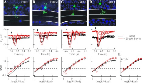
Fig. 6.
Strychnine did not influence response threshold in any of the 4 subclasses of OFF cone bipolar cells. A–D, top: type 1–4 OFF cone bipolar cells in Gnat2−/− retinas were identified on the basis of stratification of the axon terminal in the inner plexiform layer and the width of the dendritic arbor. White lines denote the limits of the inner plexiform layer, with the midpoint separating OFF from ON sublamina marked with a gray line. Middle: current-clamp recordings of flash families in the absence (black) and presence (red) of 20 μM strychnine for the photographed cells at top. Application of strychnine in all 27 OFF cone bipolar cells led to a depolarization of the cell's resting membrane potential (Vm), from −42.8 ± 1.3 mV in Ames medium to −37.6 ± 1.2 mV in Ames medium with strychnine (P = 0.0013). Bottom: SNR of flash responses plotted vs. flash strength for the cells shown at middle were fit with a saturating exponential function from which response threshold was determined (see materials and methods). E: collected data from 27 OFF cone bipolar cells across all subclasses indicate that despite the depolarization of the resting membrane potential response threshold was unaffected by the presence of strychnine and was 0.012 ± 0.002 R*/Rod in Ames medium and 0.015 ± 0.0022 R*/Rod in Ames medium with strychnine (P = 0.624).

