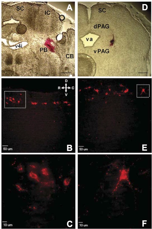
Fig. 1.
Retrograde labeling of developing lamina I neurons which project to the parabrachial nucleus (PB) or periaqueductal gray (PAG). A: section of rat brain at postnatal day (P)3 illustrating site where DiI was injected into PB at birth. SC, superior colliculus; IC, inferior colliculus; CB, cerebellum; v4i, fourth ventricle (isthmal). Scale bar, 400 μm. B: sagittal spinal cord section illustrating narrow band of retrogradely labeled neurons within lamina I at P21 after injection of DiI into PB at birth. Orientation arrows indicate dorsal (D), ventral (V), rostral (R), and caudal (C) axes. C: higher magnification of boxed region in B. D: P3 brain section demonstrating location of DiI injection into PAG at birth. dPAG, dorsal PAG; vPAG, ventral PAG; va, aqueduct. Scale bar, 400 μm. E: example of lamina I neurons fluorescently labeled at P21 after injections of DiI into PAG at birth. Same orientation as in B. F: higher magnification of boxed region in E.

