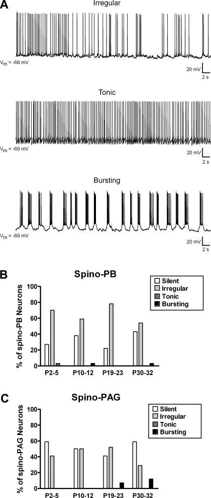
Fig. 2.
Spontaneous firing patterns in developing lamina I projection neurons. A: spino-PB and spino-PAG neurons located in lamina I of rat spinal cord were classified as irregular (exhibiting intermittent spike activity; top), tonic (continuous firing at a relatively constant frequency; middle), bursting (demonstrating rhythmic burst-firing; bottom), or silent (lack of action potential discharge; not shown). Vm, membrane potential. B and C: patterns of spontaneous activity (SA) in spino-PB (B) and spino-PAG (C) neurons at different postnatal ages, illustrating predominance of irregular spike discharge in both groups throughout development and greater overall prevalence of SA in the spino-PB population. Data for spino-PB group: P2–5: n = 33 cells from 3 rats; P10–12: n = 29 cells from 3 rats; P19–23: n = 27 cells from 4 rats; P30–32: n = 28 cells from 4 rats. Data for spino-PAG group: P2–5: n = 27 cells from 4 rats; P10–12: n = 28 cells from 4 rats; P19–23: n = 27 cells from 5 rats; P30–32: n = 17 cells from 3 rats.

