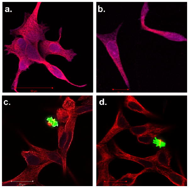
Figure 7.
Confocal microscopy images of LNCaP cells stained with PathScan apoptosis and proliferation kit. The cell were separately dosed with a) free gemcitabine, b) Et-GEM particles, c) tBu-GEM particles, and d) blank particles. Red indicates a healthy cell containing fundamental cytosolic fibers important in meiotic / mitotic chromosome alignment. Green indicates a healthy cell undergoing microtubule assembly during mitosis. Purple indicates cytoskeleton proteins and nuclear protein experiencing an apoptotic event.

