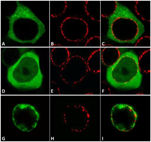
Figure 2. Localization of the Drosophila Rad9, Hus1 and Rad1 proteins in mammalian.
Human Embryonic Kidney 293 (HEK293). Confocal images of cells expressing (A) GFP-DmHus1, (D) GFP-DmRad1, and (G) GFP-DmRad9A. (B, E and H) Antibody staining of the NUP 414 protein, which recognizes several nucleoporins. (C, F and I) are merged images of (A–B), (D–E), and (G–H), respectively.

