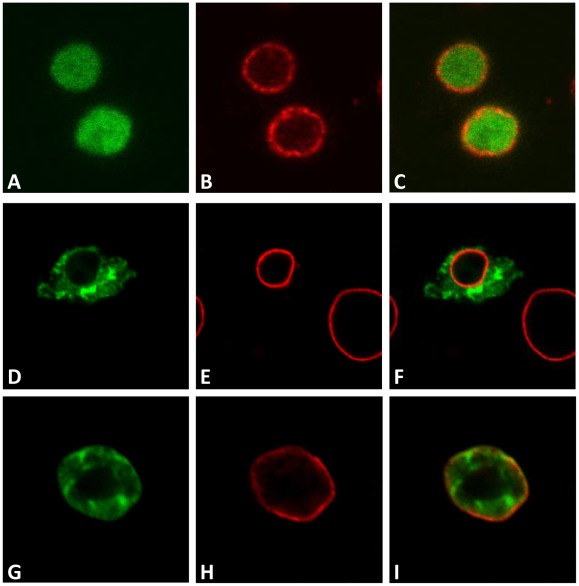
Figure 5. Identification of the DmRad9B nuclear localization signal.
Confocal images of S2R+ cells expressing DmRad9A mutated in suspected NLS sequences. (A) DmRad9B mutated at position 287 – 289 (NLS1). (D) DmRad9B mutated Position 300–302 (NLS2). (G) DmRad9B mutated Position 314–316 (NLS3). (B, E and H) stained with anti-lamin antibodies, which mark the nuclear membrane, in red. (C) Merged image of (A) and (B). (F) Merged image of (D) and (E). (I) Merged image of (G) and (H).

