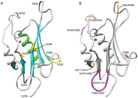
Figure 7. RNaseH fold of MuA encompassing the amino acid region E258–G396.
(A) The conserved secondary structural elements, the central β-sheet (with numbered strands 1–5) and the adjacent α-helix, are shown with light blue and light green, respectively. The DDE-motif residues are shown in yellow with exposed sidechains. Pentapeptide-insertion-tolerant sites G282, G329, and R355 are colour-coded as in Figure 4. (B) Mapping of the alignment-based insertions and deletions. The orange-coloured amino acids depict insertion sites, and the accompanying numbers identify the respective amino acids (insertions occur between the specified residues). Maximum length deletions are coloured with magenta and indicated with respective amino acid residue numbers.

