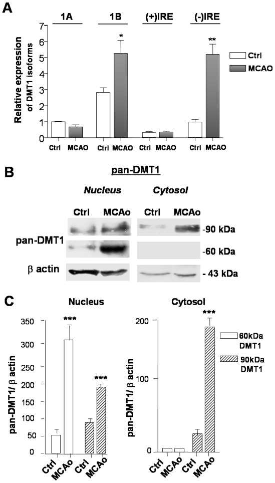
Figure 2. 1B/(−)IRE DMT1 is up-regulated in ischemic cortices of mice exposed to tMCAO.
(A) Gene expression analysis of the differentially spliced DMT1 isoforms by qRT-PCR in ischemic brain cortices of mice exposed to 1 hour transient MCAO followed by 1 hour reoxygenation compared with contralateral hemispheres (n = 3 per group). 1B/DMT1 and (−)IRE isoforms were significantly induced after MCAO. Values are expressed as fold change relative to 1A/DMT1 amplified in control (contralateral) hemisphere after normalization against β-actin cycles. Error bars represent ΔCT means±s.e.m of two independent experiments performed in triplicate, *p<0,05, **p<0,01. (B) Representative image of pan-DMT1 immunoreactivity in nuclear and cytosolic extracts from brain cortices of mice exposed to 20 min of transient MCAO or from contralateral hemisphere (n = 3 per group). Nuclear extracts were prepared 4 h after the end of the experimental condition. The pan-DMT1 reactivity increased in brain extracts of mice exposed to MCAO, both in the nuclear and in the cytosolic fractions. (C) Densitometric analysis of pan-DMT1 reactivity. Values are expressed as ratios relative to β-actin levels. Error bars represent means ± s.e.m. of three separate experiments, ***p<0.007 vs. corresponding control values.

