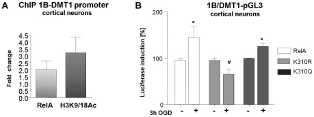
Figure 5. RelA activation of 1B/DMT1 promoter.
(A) Chromatin Immunoprecipitation assay (ChIP) for mouse1B/DMT1 proximal promoter region showed increased binding of RelA subunit and acetylation of promoter-associated histone H3(K9/18) in cortical neurons exposed to 3 hours OGD, followed by 2 hours reoxygenation. Data are normalized against total chromatin DNA used for real time PCR reaction (Input) and IgG negative control determined for each reaction, respect to control samples. (B) Primary cortical neurons at 10 DIV were co-transfected with a mouse DMT1 luciferase reporter plasmid (m1B/DMT1-pGL3) and wild-type RelA or RelA-K310R or RelA-K310Q plasmids for 24 hours. The relative luciferase activity, normalized by Renilla luciferase, was measured after 3 hours of OGD. Wild-type RelA over-expression enhanced the OGD-induced DMT1 promoter activity, as well as the acetyl-mimic RelA-K310Q construct, while RelA-K310R over-expression significantly inhibited it. Bars are means ± s.e.m of three experiments run in triplicate. *p<0.05, vs. relative Control; #p<0.05 vs. RelA OGD.

