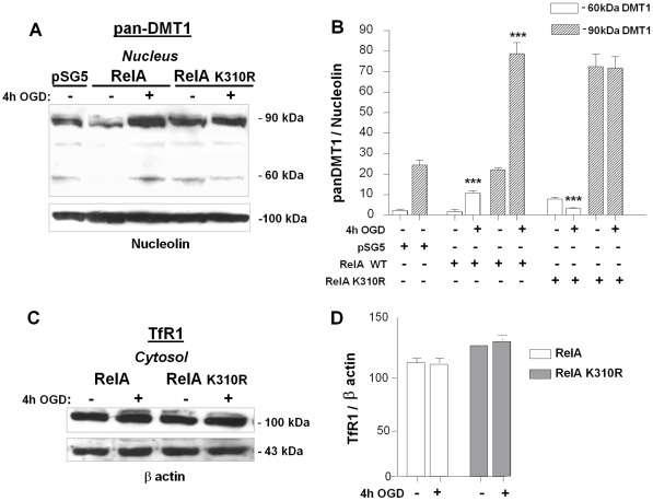
Figure 6. 1B/(−)IRE DMT1 mediates the NTBI iron transport during OGD through Lys310-acetylated RelA.
(A) Pan-DMT1 immunoreactivity in nuclear extracts from neuronal SK-N-SH cells transfected with wild-type RelA or RelA-K310R plasmids for 24 hours and further exposed to 4 hours of OGD. OGD produced a significant up-regulation of (−)IRE DMT1 protein in wild-type RelA overexpressing cells, with basal levels of DMT1 analogous to the empty vector (pSG5) transfected cells. Expression of RelA-K310R down-regulated the immature, partially glycosylated 60 kDa DMT1 isoform and failed to up-regulate the fully glycosylated 90 kDa DMT1 component. (B) Densitometric analysis of a representative pan-DMT1 immunoblots relative to nucleolin levels. Data from densitometric analysis of anti-panDMT1 antibody are expressed as as ratio relative to Nucleolin levels . Bars are the mean ± s.e.m. of three separate experiments (***p<0,0001 vs corresponding control value). (C) Immunoblot with anti-TfR antibody of cytosolic extracts from neuronal SK-N-SH cells transfected with wild-type RelA and RelA-K310R plasmids for 24 hours and exposed to 4 hours of OGD. The TfR protein level was not significantly modified in the early OGD phase in either RelA or RelA-K310R overexpressing cells. (D) Data from densitometric analysis of anti-TfR antibody immunoblots are expressed as a ratio relative to β-actin levels. Bars are means ± s.e.m. of three separate experiments.

