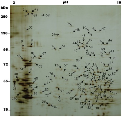
Figure 3. Representative example of 2D-gel of Chilo suppressalis larval midgut BBMV.
BBMV were separated by 2D-gel electrophoresis followed by silver staining. Positions of molecular size markers (kDa) are indicated on the side of gel. Abundant protein spots were excised (numbers 1–100), digested, and subjected to mass spectrometry analysis for identification. Spot numbers correspond to the proteins listed in Table S3.

