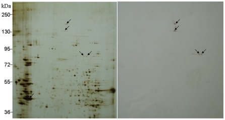
Figure 4. Blot analysis of Cry1Ac binding proteins in Chilo suppressalis midgut.
Proteins were separated by 2D-gel electrophoresis. Arrows denote positions of the Cry1Ac binding proteins. To detect Cry1Ac binding proteins, filters were probed with biotin-Cry1Ac. Positions of molecular size markers (kDa) are indicated on the side of gel.

