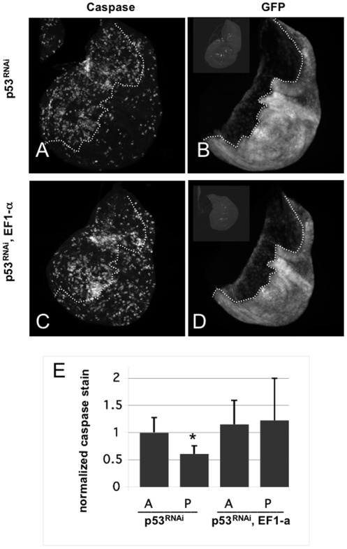
Figure 2. EF1a mutants show elevated levels of IR-induced apoptosis in a p53-depleted background.
Wing imaginal discs were dissected from 3rd instar larvae at 24 hr after exposure to 0 (−IR) or 4000R (+IR) of X-rays. Apoptosis was detected by staining with an antibody to active cleaved Caspase 3. GFP boundary is used to mark the boundary between anterior and posterior compartments. en-GAL4 is active only in the posterior compartment. (A and B) p53RNAi = en-GAL4>UAS-dsRNA against p53, UAS-GFP. Caspase stain is in (A) and GFP fluorescence is in (B). (C and D) p53RNAi, EF1-a = same as in (A) but in homozygous EF1-a mutant background. (Insets in B and D) show unirradiated control discs stained for caspase, to show little or no apoptosis in the absence of irradiation. The insets are shown with increased brightness to make disc outlines discernable. (E) Mean caspase signal in each compartment is quantified and shown normalized to the mean caspase signal of the anterior (A) compartment in p53RNAi discs (the first bar). Caspase signal in the posterior (P) compartment of the same discs are reduced significantly compared to the A compartment (p<0.001, two-tailed t-test). This is expected; the level of p53-independent apoptosis is about half of p53-dependent apoptosis at 24 hr after irradiation [8]. Caspase signal in the A compartment of ‘p53RNAi, EF1-a’ discs are not significantly different from the caspase signal in the A compartment of p53RNAi discs (p = 0.29), suggesting that reduction of EF1-a alone did not affect the level of apoptosis when p53 is present. Caspase signal in the A and P compartments of ‘p53RNAi, EF1-a’ discs are not significantly different from (p = 0.70). Caspase signal in P compartment of ‘p53RNAi, EF1-a’ discs are significantly greater than the signal in the P compartment of p53RNAi discs (p<0.05). The data are from 12 p53RNAi discs and 22 p53RNAi, EF1-a discs in two different experiments. Error bar = 1 STD.

