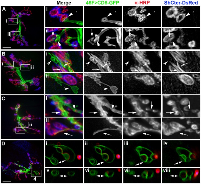
Figure 2. Glial processes at the NMJ had a wide range of morphologies.
A–D) Live NMJs with the perineurial glial membranes labeled using 46F>CD8-GFP (green), neurons immunolabeled with anti-HRP (red) and the SSR labeled using ShCter-DsRed (blue). The panels are projections of the entire NMJ (12–14 µm thick) from F3 larvae grown at 25°C. Boxed inserts (i–viii) were digitally scaled 400%. Scale bars, 15 µm. A) A NMJ where the glial processes end with blunt processes at the axon terminus just prior to the first proximal bouton (Ai, Aii; arrowheads). Glial processes also contacted discrete regions of each bouton (Aii; arrows). Glial area = 13.7 µm2. B) A NMJ where the glial processes extended across the muscle surface (Bi, Bii; concave arrowheads) or track along a channel over the bouton and SSR (Bi; arrows). Glial area = 20.7 µm2. C) A W3 NMJ where glial processes had extensive projections into the NMJ and tracked along the center of the synaptic region (Ci, Cii; arrows). Glial area = 16.85 µm2. D) A NMJ where glial processes formed node-like processes embedded in the muscle (concave arrowhead). These processes extended away from the NMJ and were not associated with boutons or SSR. Glial area = 18.6 µm2. D.i–D.iv) Inserts show the node-like glial processes (double arrowheads) embedded in the muscle imaged over a series of single 0.2 µm focal planes. D.v–D.viii) The boxed region in panel D was re-sectioned left to right (v to viii) on the YZ axis. The glial nodes (double arrowheads) were spherical and approximately 3 µm thick. The top of each panel is the approximate coverslip position.

