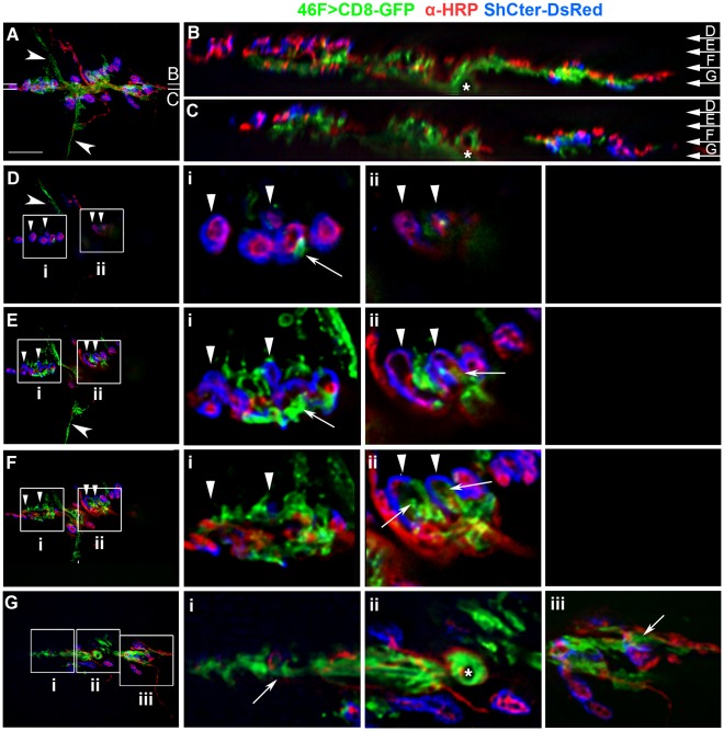
Figure 3. Perineurial glial processes interdigitated with boutons and the SSR.
A–F) A live NMJ from a F3 larva raised at 25°C. The perineurial glial processes are labeled with 46F>CD8-GFP (green), the neurons live labeled with anti-HRP (red) and the SSR labeled using ShCter-DsRed (blue). A) A projection of the NMJ (12–14 µm thick). Perineurial glial processes extended through the synapse and beyond the NMJ to contact the muscle surface (concave arrowheads) independently of neuronal or SSR structures. Glial area A = 16.7 µm2. Scale bar, 15 µm. B–C) The image stack was rotated at the points indicated in panel A by 90°. The motor axons (asterisk) entered the space between muscle 6 and 7. The GP extended along the base of the NMJ and sent projections up into the overlying bouton/SSR regions. The top (D arrow) represents the surface closest to the viscera, the bottom (G arrow) represents the layers closest to the cuticle and nerve root. D–F) Individual focal planes taken around the points indicated in panels B and C. The boxed regions were digitally scaled 200% and shown to the right (i–iii). D) A superficial focal plane showing the glial processes extending across the muscle (concave arrowhead). A few glial processes extended from below to contact the boutons/SSR (Di, Dii; arrowheads). E) A focal plane where glial processes extended across the muscle (concave arrowhead). At this level the GFP-labeled processes interdigitated with the boutons/SSR and extended between boutons (Ei, Eii; arrowheads) plus contacted the center of the synapse (Ei, Eii; arrows). F) A deeper focal plane within the middle of the NMJ. At this plane the glial processes extended to the bouton layer in the corresponding panels in E (Fi, Fii; arrowheads) or projected along the center of the synapse (Fi, Fii; arrows). G) A deep focal plane at the level of the nerve root entry between muscles 6 and 7. The motor axon entry point and the anti-HRP antibody excluded by the glial sheath are indicated (Gii; asterisk). Glial processes were more expansive at this level and extend along the length of the axons (Gi, Giii; arrows).

