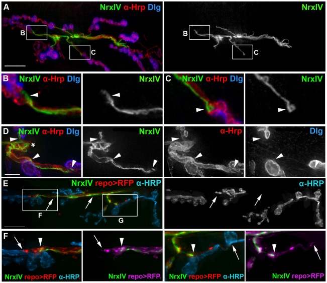
Figure 5. Neurexin IV defined the motor axon end and the septate junction barrier proximal to the synaptic boutons.
A–D) Fixed NMJ preparations where the septate junctions generated by the subperineurial glia (SPG) were labeled using Neurexin IV-GFP (NrxIV, green), the boutons and axons with anti-HRP (α-HRP, red) and the post-synaptic SSR with anti-Dlg (Dlg, blue). A 2D projection of the entire stack is shown in each panel. A) A fixed NMJ from a W3 larva with the corresponding grayscale showing the Neurexin IV-GFP. Scale bar, 15 µm. B–C) The boxed regions in panel A were digitally scaled 400% and the corresponding grayscale panels show the Neurexin IV-GFP distribution. Neurexin IV is excluded from synapses; limited to small axon branches and terminates prior to the first bouton. The septate junction termini formed blunt or tapered ends (B; arrowhead) with the occasion bulb-like structure (C; arrowhead). D) A fixed NMJ from a F3 larva in which the panels have been digitally scaled 300%. The septate junction continued along the axon from the root (asterisk) but stopped just before the proximal bouton of each branch (arrowheads). Scale bar, 5 µm. E–F) A live NMJ from a F3 larvae with the septate junctions labeled with Neurexin IV-GFP, the glia labeled using repo>CD8-RFP (red) and the neurons/boutons lived labeled using anti-HRP antibodies (blue). E) Fluorescently tagged anti-HRP antibody (α-HRP, blue) labeled the NMJ including those areas contacted by glial processes (repo>RFP, red) but the axons remain unlabeled (arrows) in the regions bounded by the septate junctions (NrxIV, green). The grayscale image shows the extent of anti-HRP antibody immunolabeling and the unlabeled axons (arrows). F, G) The boxed regions in panel E were digitally scaled 200%. Glial processes (arrows) labeled with RFP (repo>RFP) (I, J; red: Ii, Ji; magenta) extend beyond the terminus of the septate junction (NrxIV, green) (arrowheads).

