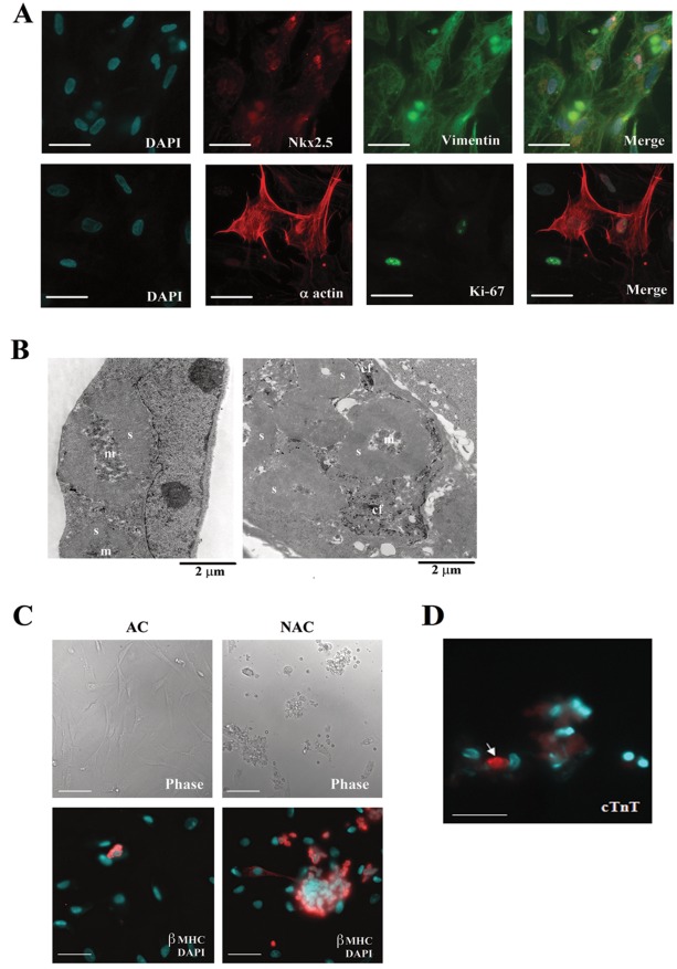
Figure 1. Cellular phenotypes in primary cultures of human fetal myocardium.
(A) Cells freshly isolated from 22 week fetal myocardium were cultured for 2 days and then immunostained with anti-nk×2.5 (red) and anti-vimentin (green) (top panel) or with anti α-actin (red) and anti ki-67 (green) (bottom panel) antibodies to determine the percentage of the cells with cardiomyocyte or fibroblast phenotypes. Nuclei were marked with DAPI staining (blue). Scale bar, 30 µm. (B) Transmission electron microscopy showing that cultures of cells freshly isolated from human fetal myocardium at day 2 contain primitive cardioblasts with nascent sarcomeres (s) and mitochondrial clusters (m) (left) and cells with the transitional features containing both nascent sarcomeres and deep invaginations containing collagen fibers (cf) (right). (C) Only a subset of cardioblasts expressed cardiac troponin T (cTnT). (D) Phase contrast images (upper panel) and fluorescent images (lower panel) showing the adherent (AC) and non-adherent (NAC) cells 2 days after isolation. Immunostaining with anti-β-MHC antibody demonstrates that non-adherent clusters consist mainly of β-MHC positive cardioblasts. Scale bar, 80 µm (for phase) and 25 µm (for fluorescent imaging).

