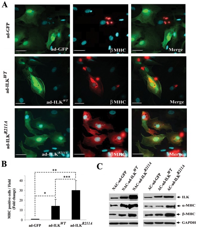
Figure 6. Over-expression of ILK in human fetal cardiac cells induces production of β-MHC.
(A) Immunofluorescent staining for β-MHC (red) in human fetal heart-derived cells (21 weeks gestation) infected with adenovirus encoding ad-ILKWT, ad-ILKR211A and ad-GFP. Nuclei were detected with DAPI staining (blue). Scale bar, 30 µm. (B) Quantification of the number of β-MHC positive cells detected by immunostaining in adherent fetal cardiomyocytes infected with adenovirus encoding ad-ILKWT, ad-ILKR211A and ad-GFP. Bar graphs represent mean values ± SD, n = 14 (random fields), *p<0.02, **p<0.001, ***p<0.03. (C) Western Blot analysis for ILK, cardiac specific α-MHC and β-MHC expression levels in adherent (AC) and non-adherent (NAC) fetal cardiac fractions infected with ad-ILKWT, ad-ILKR211A and ad-GFP. Each experiment was performed at least three times on independent samples and one representative blot is shown.

