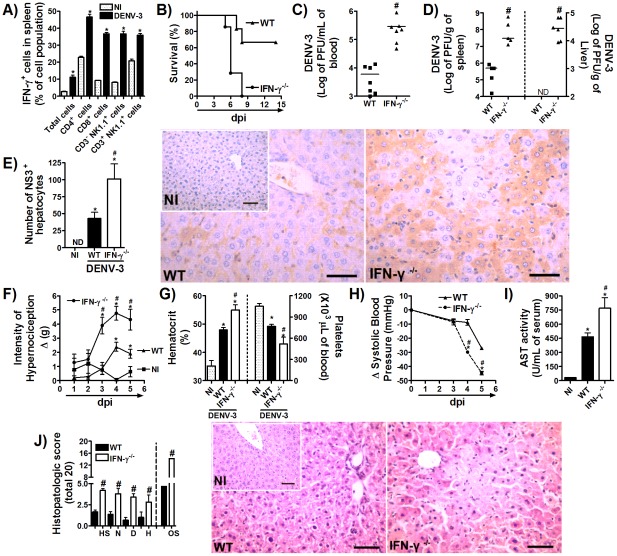
Figure 3. IFN-γ production is required for host resistance to adapted-DENV-3 primary infection.
(A) WT mice (n = 4 mice per group) were inoculated with 10LD50 (1000 PFU) of DENV-3 (i.p) and seven days later, mice were culled, and splenic cells isolated for assaying IFN-γ production by cellular staining with labeled antibodies and FACS analysis. Results are expressed as % of IFN-γ-positive cells in each population. (B) WT and IFN-γ−/− mice (n = 8 per group) were inoculated with 1LD50 (100 PFU) of DENV-3 (i.p) and lethality was evaluated every 12 hours during 14 days. Results are expressed as % of survival. In (C–J), WT and IFN-γ−/− mice (n = 6 per group) were inoculated with 1LD50 (100 PFU) of DENV-3 (i.p) and in the fifth day of infection mice were culled and blood and tissues were collected for the following analysis: (C–D) Viral loads were recovered from the blood (C), spleen and liver (D, left and right panels), respectively. Results are shown as the log of PFU per mL of blood or per g of tissue. (E) Serial sections from each liver were stained with anti-DV NS3 antibody E1D8 (NS3) or an isotype control mouse IgG2a (IgG2a data not shown), and multiple sections of each tissue type were thoroughly examined for staining. Positive staining for NS3 is brown while hematoxylin counterstain is blue. Results are expressed as number of NS3-positive hepatocytes. (F) Mechanical hypernociception was assessed daily. Results are shown as the difference between the force (g) necessary to induce dorsal flexion of tibio-tarsal joint, followed by paw withdraw, before and after DENV-3 inoculation. In (G), hematocrit was shown as % volume occupied by red blood cells (left panel) and the number of platelets was shown as platelets ×103/µl of blood (right panel). (H) Changes in Systolic blood pressure from baseline until day 5 after infection expressed as Δ of blood pressure in mmHg. In (I), AST activity determination in plasma, shown as U/dL of plasma. (J) shows semi-quantitative analysis of hepatic damage (histopathological analysis performed as modified from Paes et al, 2009) and Hematoxylin & Eosin staining of liver sections of control and WT and IFN-γ−/− DENV-3-infected mice, five days after infection. Scale bars - 400 µm. The images presented are representative of an animal on the fifth day of infection. All results are expressed as mean ± SEM (except for C–D, expressed as median) and are representative of at least two experiments. * for P<0.05 when compared to control uninfected mice. # fo P,0.05 when compared to WT infected mice. 10 LD50 corresponds to 1000 PFU of adapted-DENV-3. 1LD50 corresponds to 100 PFU of adapted-DENV-3. ND – not detectable. NI- Not-infected. dpi – days post-infection. HS – hepatocyte swelling. N – necrosis. D – degeneration. H – hemorrhage. OS – Overall Score.

