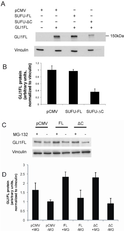
Figure 6. SUFU-ΔC but not SUFU-FL reduces GLI1FL protein levels. A.
, GLI1FL was introduced into Hek293 cells together with either SUFU-FL or SUFU-ΔC FLAG-tagged expression constructs, as indicated. Soluble protein fractions were run on a SDS-PAGE gel, followed by Western blotting, and detected by a GLI1 antibody. Vinculin staining was used as a loading control. B, Quantification of average GLI1FL protein levels in this and in an additional repeat experiment. Note that SUFU-ΔC but not SUFU-FL elicits a reduction of GLI1FL protein. C, SUFU-FL (FL) or SUFU-ΔC (ΔC) was introduced into RMS13 cells, with or without 10 µM MG-132 treatment for 6 hours. Soluble protein fractions were run on a SDS-PAGE gel, followed by Western blotting, and detected by a GLI1 antibody. Vinculin staining was used as a loading control. D, Quantification of average GLI1FL protein levels in this and in an additional repeat experiment. Note that SUFU-ΔC but not SUFU-FL elicits a detectable reduction of GLI1FL protein and that MG-132 treatment confers an increase in GLI1FL protein.

