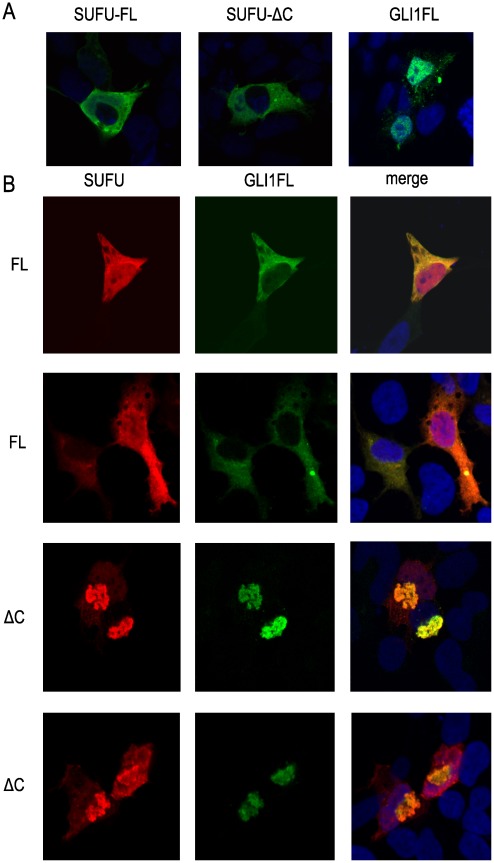
Figure 7. Subcellular localization of SUFU-FL and SUFU-ΔC in Hek293 cells.
The transfected SUFU and/or GLI1FL constructs were detected using Myc and FLAG antibodies by confocal microscopy. A, Individual transfections of Myc-tagged SUFU-FL or SUFU-ΔC and FLAG-tagged GLI1FL constructs. B, Co-transfections of GLI1FL with the SUFU-FL (FL) or the SUFU-ΔC (ΔC) construct. Two representative images from each co-transfection are shown. The nuclei are stained with the marker DRAQ5 (blue signal). Note that GLI1FL co-localizes with SUFU-ΔC in aggregate/clump structures in the cytoplasm.

