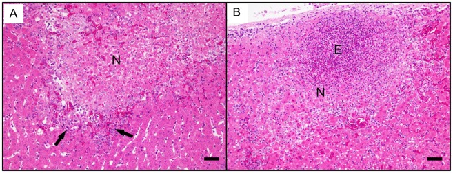
Figure 7. The effects of vaccination with rFhGST-S1 or Quil A.
A) Photomicrograph of the liver from the Quil A immunised group showing an area of coagulative necrosis (N) surrounded by scarce inflammatory infiltration (arrows) with occasional eosinophils. B) Photomicrograph of the liver from the rFhGST-S1 immunised group showing a coagulative necrotic area (N) associated to numerous eosinophils (E). Both images haematoxylin and eosin stained. Both bar represent 100 µm.

