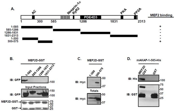
Figure 3. MEF2 directly binds amino acids 300-375 on mAKAP.
(A) Schematic diagram of the mAKAP fragments used for pulldown assays. (B) MEF2D-GST beads were incubated with HEK293 cell lysates transfected with the indicated mAKAP fragments. After extensive washing, association of the mAKAP fragments was visualized by immunoblot using a GFP antibody. Total protein expression is shown in the middle panel while protein stains of the MEF2D-GST fusion protein is shown in the lower panel. n=3. (C) HEK293 cells were transfected with myc-tagged mAKAP fragments encompassing amino acids 1-300, or 300-575. Cell lysate was isolated and subjected to pulldown assay using GST beads charged with MEF2D. Association of the mAKAP fragment was demonstrated by immunoblot using a myc antibody. Total protein expression is shown in the lower panel. n=3. (D) Bacterially purified His-tagged mAKAP-1-585 was incubated with either MEF2D-GST beads or GST control, and association of mAKAP was visualized by immunoblot using an antibody that recognized the His tag on the mAKAP fragment. The lower panel is a western blot of the GST-fusion proteins used. n=4.

