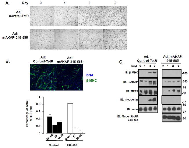
Figure 7. MEF2 tethering to mAKAP is required for efficient myogenic differentiation.
(A) C2C12 cells infected with either a control adenovirus (top row) or adenovirus expressing myc-tagged mAKAP-245-585 were imaged over four days to monitor the development of myotubes. Scale bar = 20 μm. n=3. (B) C2C12 cells were treated as describe in A. After three days in differention media, the cells were stained with MHC and Hoechst. The number of nuclei per β-MHC positive cell was determined from three separate experiments. A total of 304 cells were counted from the 245-585 expressing cells while 367 cells were counted from the control. (C) Immunoblot analysis of differentiation markers from lysates prepared from C2C12 cells infected with either control adenovirus or adenovirus expressing myc-mAKAP-245-585 over the four days of imaging described in (A). n=3.

