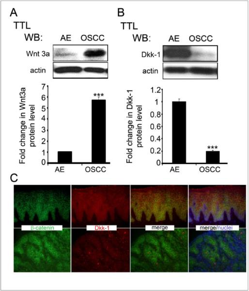
Figure 3. Upregulation of canonical Wnt in OSCC is associated with decreased Dkk-1 and elevated Wnt3a levels.

(A) Western blot of Dkk-1 expression in AE and OSCC. Bargraph, Fold change in Dkk-1 levels after normalization to actin (***p< 0.001). (B) Western blot of Wnt3a expression in AE and OSCC. Bargraph, Fold change in Wnt3a levels in OSCC in comparison to AE after normalization to actin (**p< 0.01). (C) Immunofluorescence staining of Dkk-1 and b-catenin in AE and OSCC. Size bar: 50 μm. All results represent one of three independent experiments
