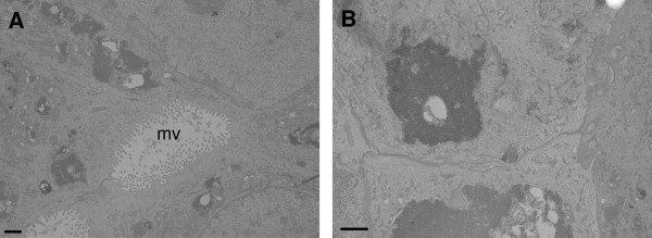
Figure 7.
The effect of T-2 toxin on the morphology of differentiated IPEC-J2 cells. Transmission electron micrographs of differentiated IPEC-J2 cells fixed 24 h after exposure to (A) control medium or (B) 5 ng/mL T-2 toxin. These pictures serve as a representative for a confluent monolayer of IPEC-J2 cells and no differences were seen on the ultrastructure of T-2 toxin (5 ng/mL) treated IPEC-J2 cells in comparison to untreated cells. Scale bar = 1 μM; mv = microvilli.

