
Figure 1.
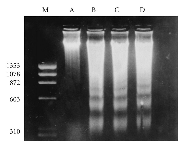
Morphine induces apoptosis of human PBMCs. PBMCs were cultured alone or with different concentrations of morphine for 60 h. Total DNA was extracted and electrophoresed on a 1.8% agarose gel in the presence of ethidium bromide. Arrows indicate fragmented DNA. Lane A, control culture; lanes B and C, cells treated with morphine at concentrations of 10−7 and 10−9 M respectively; lane D, cells treated with cortisol at 0.2 mg/mL (positive control); lane M, molecular weight marker. Copyright © 1997, American Society for Microbiology. This figure is reproduced from the original article published in Clinical and diagnostic laboratory immunology, Nair et al. [35].