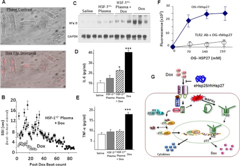
Fig. 8.
Dox-induced toxicity and protection by HSF-1(+/−) plasma in adult cardiomyocytes. A, phase-contrast and Dox fluorescence (Flu.) (merged) of Dox-treated myocytes. B, quantitative plots of BBI for cardiomyocytes after addition of Dox in the culture medium (n = 6). C, representative Western blots of NF-κB in the nuclear extracts from all the four groups. D and E, IL-6 and TNF-α determined in all the four groups (***, p < 0.001; *, p < 0.01). F, fluorescence measured in OG-hHsp27 treated and TLR2 antibody (Ab) + OG-hHsp27-treated cardiomyocytes. G, schematic illustration of mechanism of cardioprotection by eHsp25 against Dox in the heart. Dox binding to TLR2 activates the NF-κB-related pathway of apoptosis, as well as MAPKs. This MAPK activation may also be assisted by intracellular redox reaction of Dox to activate HSF-1 and to induce Hsps, particularly Hsp25, which in turn transactivates p53 and expression of Bax (Vedam et al., 2010). If there is a higher level of Hsp25 in the circulation, it can competitively bind to the TLR2 receptors, antagonizing Dox to inhibit Dox-induced apoptotic signaling.

