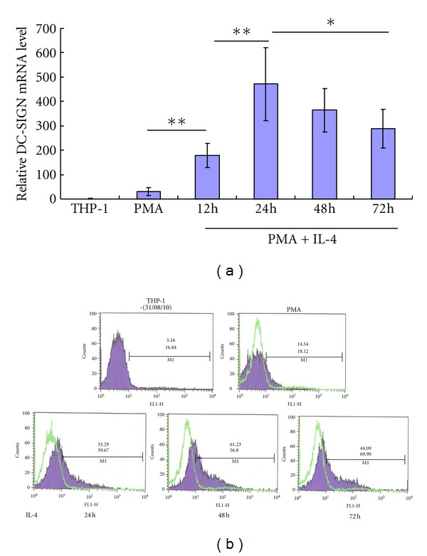
Figure 1.

IL-4-induced high expression of DC-SIGN on THP-1 cells over time. THP-1 cells were treated with or without PMA for 24 hours, or with PMA for 24 hours and IL-4 for up to 72 hours. (a), the quantitative analysis of the level of DC-SIGN mRNA in differentiated THP-1 cells by PMA and IL-4 by SYBR Green real-time PCR. (b), analysis of induced DC-SIGN expression on surface of differentiated THP-1 cells by flow cytometry. The percentage of positive cells (top number) and mean fluorescence intensity (figure below) are shown. *means P < 0.05; **means P < 0.01.
