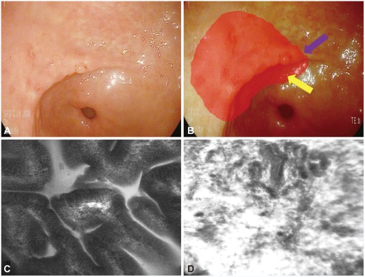
Fig. 11.
Tumor margin delineation with confocal laser endomicroscopy (CLE). (A) Slightly depressed lesion is noted and previous forceps biopsy show signet ring cell adenocarcinoma. (B) It is not easily delineated for the exact tumor margin (red circle). (C) CLE help the delineation of the tumor margin with the determination of in vivo histology (normal mucosal surface; purple arrow in B). (D) Tumor mucosal surface (yellow arrow in B).

