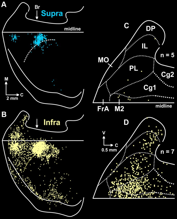
Figure 4.

Cortical maps from animals with fifth-order labeling. A, Infected neurons in supragranular layers (Layer IV and above; Supra, blue). B, Infected neurons in infragranular layers (Layers V and VI; Infra, yellow). Both maps are from the same representative animal. C, Composite map of fourth-order neurons found in cortical areas on the medial wall of the hemisphere (n = 5). Dashed lines indicate the cytoarchitectonic borders of specific regions. The figure is an enlarged view of the medial wall data shown in Figure 2B. D, Composite map of labeled infragranular neurons in the medial wall of the hemisphere. To create this figure, we overlapped the maps of labeled infragranular neurons in animals with fifth-order labeling (n = 7; for details see Materials and Methods, Analytic procedures). Each square represents a single labeled neuron. Br, bregma; C, caudal; Cg1, cingulate cortex area 1; Cg2, cingulate cortex area 2; DP, dorsal peduncular cortex; FrA, frontal association cortex; IL, infralimbic cortex; M, medial; midline, midline of the hemisphere; MO, medial orbital cortex; M2, secondary motor cortex; PL, prelimbic cortex; V, ventral.
