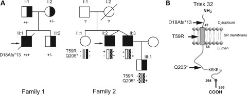
Figure 1.
Triadin mutations in two CPVT families. (A) Pedigrees of the two CPVT families. Filled squares indicated affected individuals and half filled symbols individuals heterozygous for a mutation. The proband in each Family is indicated by the black arrow. The genotype for each individual concerning the identified mutation is indicated as follow: ‘+’ in the presence of the mutation and ‘−’ in the absence of the mutation. (B) Membrane topology of cardiac isoform of triadin, Trisk 32, in the sarcoplasmic reticulum membrane with the localization of the three identified mutations. The ‘KEKE’ region is the interaction domain with RyR2 and CSQ2. Trisk 32 has a C-terminal specific region from amino acid 264–286.

