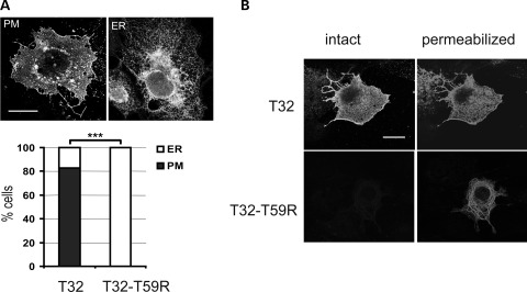
Figure 2.
Trisk 32 and Trisk 32-T59R cell localizations are different. (A) Trisk 32-T59R is located only at the endoplasmic reticulum, whereas Trisk 32 is mainly at the plasma membrane. WT Trisk 32 (T32) and mutant Trisk 32-T59R (T32–T59R) were transfected in COS-7 cells, and their localization analyzed by immunofluorescent labeling. Typical Trisk 32 labeling at the plasma membrane (PM) or in the ER are shown. Bar: 20 µm. The histogram shows the percentage of transfected cells exhibiting PM (closed square) or ER (open square) labeling, for a total of 500 transfected cells from three different experiments. ***P < 0.001, Fisher's test comparison of T32- vs. T32–T59R-transfected cells. (B) Immunofluorescent labeling on intact or permeabilized cells shows intracellular retention of Trisk 32-T59R. Transfected cells were fixed without permeabilization and stained with an antibody directed against the C-terminal end of Trisk 32, which is extracellular when the protein is in the plasma membrane (left panels, ‘intact’). Afterwards, cells were permeabilized and stained with an antibody directed against the N-terminal end of Trisk 32, which is cytosolic when the protein is in the plasma membrane or in the reticulum membrane (right panels, ‘permeabilized’). Bar: 20 µm.

