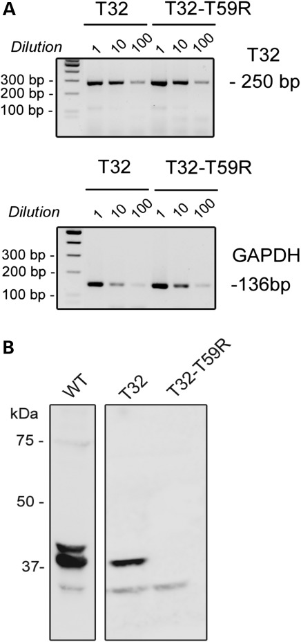
Figure 4.
Stability analyses of transcripts and proteins after in vivo expression in triadin KO mice. (A) The mutant transcript is present at similar levels as the WT one. Messenger RNAs were purified from isolated cardiomyocytes of transduced mice. After reverse transcription, the cDNAs were either directly used or diluted 10 or 100 times before the PCR amplification of Trisk 32 (T32) and GAPDH. PCR products for Trisk 32 and GAPDH amplification were expected, respectively, at 250 and 136 bp. The PCR primers for GAPDH were designed to co-amplify possible contaminating genomic DNA as a 220 bp fragment, which was not observed. (B) The mutant protein Trisk 32-T59R was not detectable. Western blot analysis of 100 µg of cardiomyocytes homogenates from control WT mouse, with mouse-specific anti-Trisk 32 antibody (first lane), or 100 µg of cardiomyocytes from a Trisk 32-transduced mouse (second lane), or Trisk 32-T59R-transduced mouse (third lane) with rat-specific anti-Trisk 32 antibody. Trisk 32 appears as multiple bands (2/3) centered on 37 kDa, the higher bands, the glycosylated forms of Trisk 32, being in lower amount and visible in correlation with the intensity of the signal, as described before (6,20).

