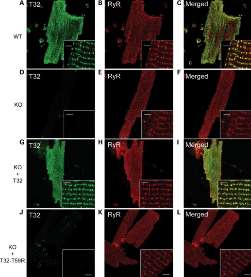
Figure 5.
Immunofluorescent analysis of Trisk 32 in isolated cardiomyocytes. Cardiomyocytes were isolated from a WT mouse, a triadin KO mouse (KO), a triadin KO mouse transduced with rat Trisk 32 (KO + T32) and a triadin KO mouse transduced with rat Trisk 32-T59R (KO + T32-T59R). They were labeled with antibodies against mouse Trisk 32 and RyR (A–F) or against rat Trisk 32 and RyR (G–L). In all cardiomyocytes, RyR labeling is typical of a dyad labeling showing aligned rows of dots, as observed in the inserts. Bar: 2 µm.

