
Figure 8.
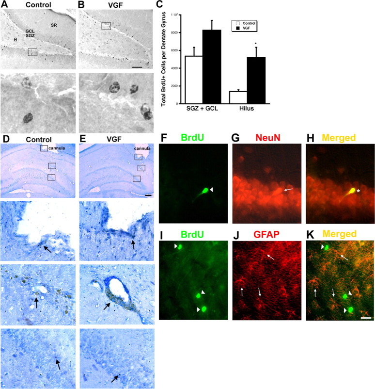
VGF-induced BrdU+ cells survive to 21 d and differentiate primarily into neurons. A–C, VGF-induced BrdU+ cells survive to 21 d. A, B, Saline (A) or VGF (4 μg) (B) was infused daily via a cannula into the hippocampi of adult male rats for 7 d followed by a single BrdU injection (100 mg/kg). The animals were killed 21 d later. BrdU+ cells (black) are visible in the subdivisions of the dentate gyrus including the hilus (H), subgranular zone (SGZ), granule cell layer (GCL), and stratum radiatum (SR) in coronal sections. Scale bar, 80 μm. Enlarged images of boxed regions are shown below. C, Quantitation of total BrdU+ cells in the various regions of the dentate gyrus. The bars represent average BrdU+ cells ± SE (n = 8). *p < 0.05, t test. D, E, Repeated infusions of saline or VGF do not cause damage to the dentate gyrus. Saline (D) or VGF (4 μg) (E) was infused daily via a cannula into the hippocampi of adult male rats for 7 d. The animals were killed 21 d later and processed for cleaved caspase-3 immunohistochemistry. Location of guide cannula is shown at low magnification (arrowhead). Scale bar, 150 μm. Enlarged images of boxed regions showing tip of cannula, site of infusion, and dentate gyrus, respectively, are shown below. Caspase-3-positive cells are indicated by arrows. F–H, BrdU+ cells that survive primarily express neuronal markers. F, BrdU+ cells (green; arrowhead). G, NeuN+ cells (red; arrow). H, Merged image showing colocalization (yellow; asterisk). I–K, BrdU+ cells that survive do not express glia markers. I, BrdU+ cells (green; arrowheads). J, GFAP+ cells (red; arrows). K, Merged image showing no double-labeled cells (yellow). Scale bar, 20 μm.