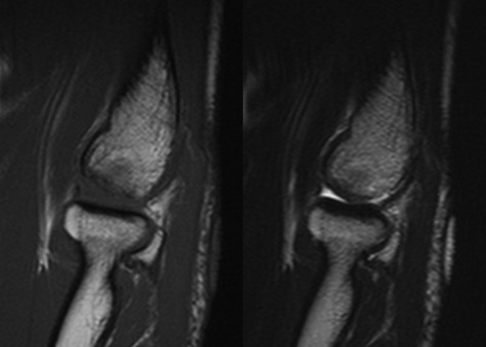
Fig. 5.
Case 11. MRI scans made twelve months postoperatively. A T1-weighted sagittal image (left) shows well-revascularized subchondral bone in the graft area, and congruent articular contours are apparent on a T2-weighted sagittal image (right). The patient returned to sporting activity as a Judo athlete within one year.

